We gladly announce the commencement of the first Fan Beam Dual Energy X-Ray Absorptiometry (DEXA) Scan in the region.
This is our sincere effort to provide state-of-the-art technology for assessing Bone Mineral Density (BMD).
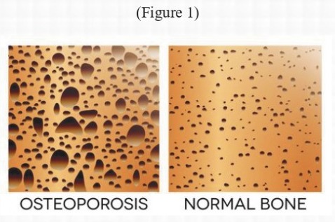
Osteoporosis is the most common metabolic bone disorder characterized by reduced bone density
with or without fragility fractures. Compromised bone strength increases fracture risk.
Though commonly seen in post-menopausal women, it is increasingly prevalent in elderly men.
After age 50, lifetime risk of osteoporotic fractures (hip, spine, wrist combined):
- 40% in females
- 13% in males
Most patients remain asymptomatic until fracture occurs. Hence, early screening is critical.
Aging and gonadal hormone deficiency disturb the bone remodeling balance, causing:
- Increased bone resorption (bone loss)
- Reduced bone formation
- Resulting in weak bones of reduced density
Peak Bone Mass & Prevention
Genetic factors, adequate Calcium & VitaminD intake, and regular exercise during childhood
determine peak bone mass.
Higher peak bone mass + active lifestyle = Lower risk of osteoporosis.
WHO Diagnostic Criteria (Based on T-Score)
The World Health Organization (WHO) defines osteoporosis based on
Bone Mineral Density (BMD), measured by DEXA scan.
T-Score compares a patient’s bone density with that of a healthy young adult of the same gender.
| T-Score | Diagnosis |
|---|---|
| At or above -1 SD | Normal |
| Between -1 and -2.5 SD | Osteopenia (Increased Risk) |
| At or below -2.5 SD | Osteoporosis (With or Without Fragility Fracture) |
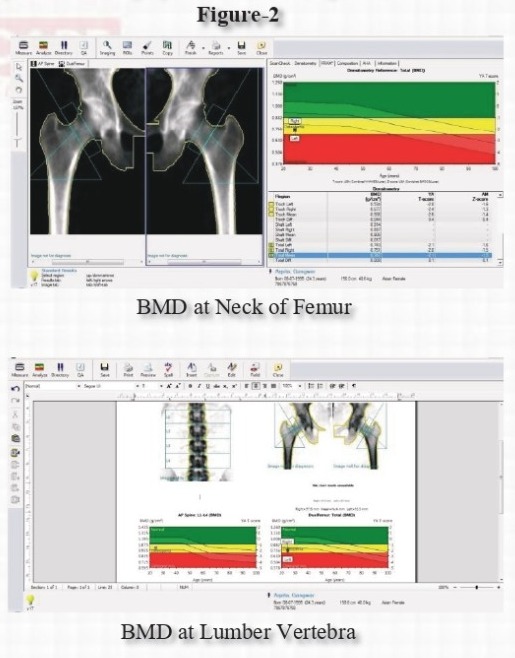
Bone density is measured at:
- Lumbar Vertebrae
- Bilateral Femoral Regions
The results are plotted graphically as shown below:
Classification of Osteoporosis
- A. Primary
- B. Secondary
- C. Idiopathic
A) Primary Osteoporosis
- Post-menopausal – Usually vertebral crush fractures.
- Senile – Older men & women; commonly associated with hip fractures.
B) Secondary Osteoporosis
- 1. Immobilization: Prolonged bed rest or chronic illness.
- 2. Endocrine Disorders: Hypogonadism, Hyperparathyroidism, Thyrotoxicosis, Cushing’s syndrome, Acromegaly, Prolactinomas etc.
- 3. Gastrointestinal Disorders: Malnutrition, Malabsorption, Hepato-biliary diseases (Primary Biliary Cirrhosis).
- 4. Rheumatological Diseases: Rheumatoid Arthritis, Ankylosing spondylitis, Marfan’s syndrome etc.
- 5. Hematological Disorders: Multiple Myeloma, Leukemia, Lymphoma, Sickle Cell Disease, Thalassemia, Gaucher’s disease etc.
- 6. Drug Induced: Steroids, Cytotoxics, Anticonvulsants, Lithium, Heparin, Excess Thyroxine therapy.
- 7. Renal Diseases: Chronic Renal Failure.
Major causes: Primary osteoporosis, Hypogonadism & Steroid-induced osteoporosis.
C) Idiopathic Osteoporosis
Osteoporosis occurring without identifiable secondary cause.
Risk Factors
- Poor Nutrition
- Low Body Weight / BMI
- Early Menopause (Before 45 years)
- Prolonged Pre-menopause Amenorrhea
- Previous Fragility Fractures
- Inadequate Physical Exercise
- Immobility
- Excessive Alcohol & Smoking
- Low Calcium Intake
Screening
Screening is recommended for post-menopausal women above 65 years and men above 70 years.
Patients on steroid therapy (≥5 mg/day for >12 weeks) should also undergo screening.
X-Ray detects osteoporosis only after 30% bone loss.
DEXA Scan (Dual Energy X-ray Absorptiometry) is the Gold Standard.
DEXA classifies patients into:
Normal, Osteopenia, and Osteoporosis.
It is sensitive, specific, reproducible, and useful for yearly follow-up.
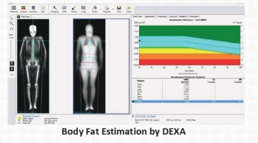
Other Uses of DEXA Scan
- Total Body Fat Estimation in overweight & obese patients.
- Used by body builders & fitness-conscious individuals.